Each person’s body contains more than 40billions of the thinnest blood vessels - capillaries. Unfortunately, even despite their importance in the distribution of oxygen in cells, the mechanism of their internal work for the most part remains a mystery to scientists. A group of researchers from Northwestern University (USA) has developed a new tool for visualizing blood flow through these tiny blood vessels, and very soon we will be able to learn much more about capillaries, writes Futurism portal.

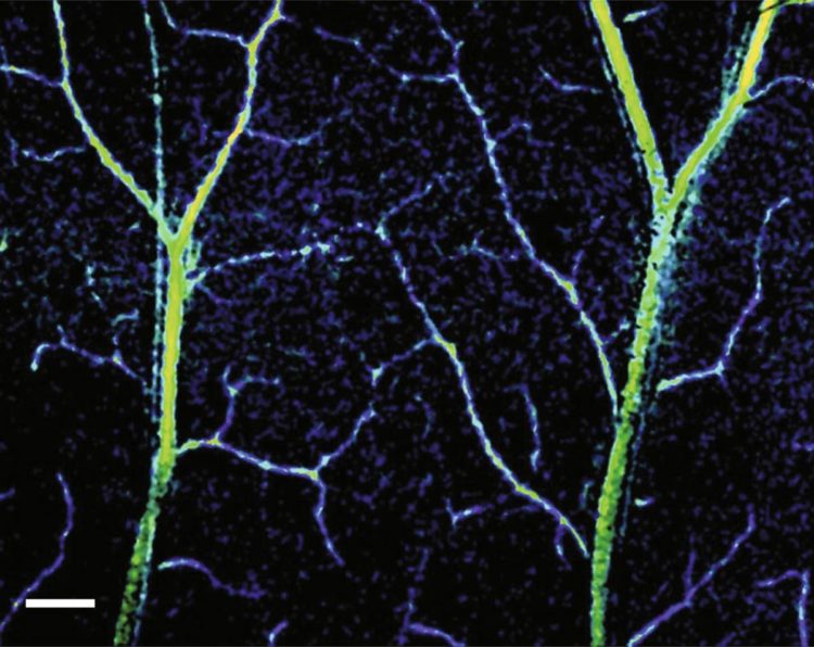
Using a method calledWith spectral contrast optical coherence tomography (spectral contrast optical coherence tomography angiography, SC-OCTA), a team of scientists was able to get an unprecedented opportunity to look at capillary blood flow. Their study, published in the journal Light: Science & Applications, can have far-reaching implications both for medicine in general and for the understanding of the human body in particular.
Researchers and physicians have long been able topeering inside large blood vessels with ultrasound. However, ultrasound does not work in all cases. For example, it is completely ineffective if you want to consider a vessel in which there is no movement of blood. If we speak directly about capillaries, a small and completely absent blood flow through capillary channels with a diameter of 5-10 micrometers can lead to poor oxygen circulation, which, in turn, can cause a variety of effects, ranging from mild headaches to heart failure .
"Despite the presence of good blood flow inarteries, you may have problems with oxygenation of the tissues if there is no proper microvasculature, ”says study leader Vadim Bakman
The advantage of the SC-OCTA method is thatit does not need the presence of blood flow in the capillaries to see a clear picture of what is happening. This can significantly simplify the task of doctors in determining what caused poor blood flow through the capillary, and in a timely manner to prevent problems associated with low oxygen levels. The Backman team notes that the method they developed uses only light and it requires the use of contrasting dyes or harmful radiation, which are usually used with traditional imaging techniques, for example, with the same ultrasound. At the same time, SC-OCTA allows accurate visualization at a depth of just 1 millimeter. Unlike the same method of ultrasound scanning, which allows you to "dive" a few centimeters, this represents a rather significant disadvantage, the researchers agree, but they immediately add that they are already working on how to increase the penetrating ability of SC-OCTA.
“This is a progressive attempt to bring the smallest blood vessel imaging technology to a new level, able to offer more complete and functional information,” says Beckman.
Scientists believe that the ability to detect evenminor changes in the structure of the capillaries will help doctors detect in advance the development of cancer or diseases of the cardiovascular system. In addition, the new method can help in ongoing research on organelles aimed at understanding how these organs react to the development of various diseases.
You can discuss the development in our Telegram-chat.
Related articles
 Announcement. Motorola Edge 40 Pro is the European flagship twin
- Sales of smartphones in Russia decreased by 26%
- Enthusiast has created a prototype of the Cyberquad ATV designed for Elon Musk's Cybertruck pickup truck (video)
- Nintendo intends to maintain a minimal presence in the Russian market
- Why does a person need air and how do lungs work?
- The largest family tree has shown where humanity originated
- AI startup brings the dead to life in the form of a hologram
- Elon Musk's company chipped a monkey and taught it to play on a computer using the power of thought